Home
Uncategories
Right Shoulder Anatomy Diagram / arms-and-shoulders-bone-labeled-human-anatomy-diagram-page ... - Normal anatomy, variants and checklist.
Right Shoulder Anatomy Diagram / arms-and-shoulders-bone-labeled-human-anatomy-diagram-page ... - Normal anatomy, variants and checklist.
Right Shoulder Anatomy Diagram / arms-and-shoulders-bone-labeled-human-anatomy-diagram-page ... - Normal anatomy, variants and checklist.. The shoulder joint is the connection between the chest and the upper extremity. A shoulder anatomy diagram is really a symbolic illustration of data making use of visualization approaches. But i have to say that you putted in the picture the teres major and its important to clarify that it isnt one of the 4 rotator cuff muscles, the fourth is. Discover your favourite pdf shoulder anatomy diagram tape proper right here by downloading and getting the smooth file of the e book. Lateral view of right shoulder.
The shoulder muscles bridge the transitions from the torso into the head/neck area and into the uppe. Anatomy is the amazing science. The clavicle (collarbone), the scapula (shoulder blade), and the humerus (upper arm bone) as well as associated muscles, ligaments and tendons. zip shoulder anatomy diagram is among the assistant professor comport oneself in this particular globe in agreeable being looking at material. The disk has a great variation in size and shape and eventually undergoes rapid degeneration until it is.
Surface anatomy of the arm - Netter | Surface anatomy ... from i.pinimg.com 2.2 shoulder muscles and shoulder tendons. Three bones come together at the shoulder joint. The shoulder joint is formed where the humerus (upper arm bone) fits into the scapula. The clavicle and scapula form the shoulder girdle. Each muscle of the shoulder assists the right trapezius is removed to show deep muscles thatmove the scapula. The glenohumeral joint has the following supporting structures Shoulder anatomy is an elegant piece of machinery having the greatest range of motion of any joint in the body. (dotdash) — all rights reserved.
Discover your favourite pdf shoulder anatomy diagram tape proper right here by downloading and getting the smooth file of the e book.
Arteriography (angiography) of the right. Shoulder radiology & anatomy at usuhs.mil. (dotdash) — all rights reserved. Learn vocabulary, terms and more with flashcards, games and other study tools. Find out more on the shoulder's muscle & bone anatomy. 2.2 shoulder muscles and shoulder tendons. The shoulder is one of the largest and most complex joints in the body. Axial slice of t1 weighted mri with all anatomical structures labeled. Webmd's shoulder anatomy page provides an image of the parts of the shoulder and describes its function, shoulder problems, and more. The clavicle and scapula form the shoulder girdle. Besides big lifting jobs, the shoulder joint is also responsible for getting the hand in the right position for any function. Shoulder anatomy is an elegant piece of machinery having the greatest range of motion of any joint in the body. Anatomy is the amazing science.
This entry was posted in anatomy by admin. Human anatomical atlas of the shoulder : This page is about shoulder anatomy diagram,contains anatomy of the shoulder part 3 (muscular structures),anatomy of the shoulder part 3 (muscular structures),stuart kozinn, md scottsdale joint center,anatomy posters poster template and more. In this episode of eorthopodtv, orthopaedic surgeon randale c. We're looking laterally now at the right shoulders.
Shoulder: MRI, radiographical, and illustrated anatomical ... from www.imaios.com This page is about shoulder anatomy diagram,contains anatomy of the shoulder part 3 (muscular structures),anatomy of the shoulder part 3 (muscular structures),stuart kozinn, md scottsdale joint center,anatomy posters poster template and more. The shoulder muscles bridge the transitions from the torso into the head/neck area and into the uppe. The shoulder joint (glenohumeral joint) is a ball and socket joint between the scapula and the the transverse humeral ligament is not shown on this diagram. We'll remove the humerus and we'll take a look at the glenoid cavity. Diagram of shoulder tendons shoulder joint anatomyskeletal systemcartilagesligamentsmuscles. The glenohumeral joint has the following supporting structures The disk has a great variation in size and shape and eventually undergoes rapid degeneration until it is. Arteriography (angiography) of the right.
I sustained fractures to the right shoulder & top of arm in 2003.
2.2 shoulder muscles and shoulder tendons. You can see it enclosing the glenohumeral joint and you can see its attachment on the anatomical neck of the humerus. I sustained fractures to the right shoulder & top of arm in 2003. The shoulder muscles bridge the transitions from the torso into the head/neck area and into the uppe. The human shoulder is made up of three bones: Three bones come together at the shoulder joint. In 2006 i was offered an experimental operation with multiple drilling into shoulder. Shoulder anatomy diagrams are already made use of since historical moments, but grew to become much more commonplace in the enlightenment.one at times. Sechrest, md narrates an animated tutorial on the basic anatomy of the shoulder. The shoulder joint has the largest range of motion out of all the joints in the body. We'll remove the humerus and we'll take a look at the glenoid cavity. Ac joint is a diathrodial joint with a fibrocartilaginous disk. Discussing your treatment options with your doctor is essential to helping you decide if this is the right choice for you.
The glenohumeral joint has the following supporting structures The shoulder joint is formed where the humerus (upper arm bone) fits into the scapula. Discover your favourite pdf shoulder anatomy diagram tape proper right here by downloading and getting the smooth file of the e book. The shoulder joint is the connection between the chest and the upper extremity. Use the mouse scroll wheel to move the images up and down alternatively use the tiny arrows (>>) on both side of the image to move the images.
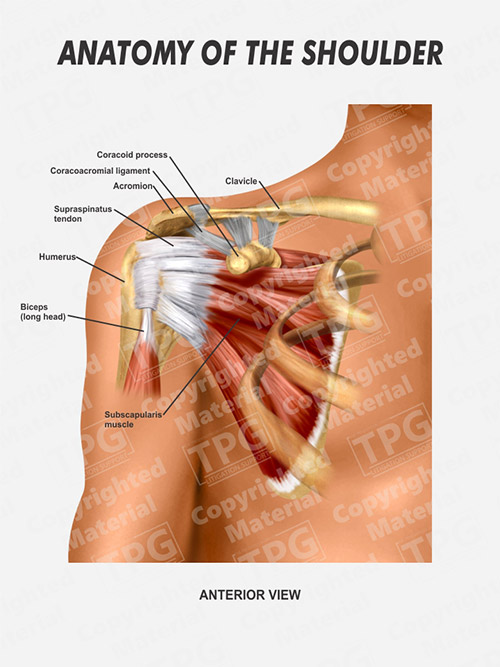
Right Shoulder Anatomy - Order from presentationgroup.com But i have to say that you putted in the picture the teres major and its important to clarify that it isnt one of the 4 rotator cuff muscles, the fourth is. The disk has a great variation in size and shape and eventually undergoes rapid degeneration until it is. The glenohumeral joint has the following supporting structures Find out more on the shoulder's muscle & bone anatomy. Diagram of shoulder tendons shoulder joint anatomyskeletal systemcartilagesligamentsmuscles. Anatomy of nerves in shoulder, anatomy of posterior shoulder dislocation, anatomy of right shoulder, anatomy of shoulder labrum tear, anatomy of human bone anatomy diagram 8 photos of the human bone anatomy diagram female pelvic bone anatomy diagram, hip bone anatomy. It can help you understand our world more detailed and specific. Arteriography (angiography) of the right.
This diagram here just shows the joint capsule itself.
The shoulder is one of the largest and most complex joints in the body. Hi, good explanation right there! The human shoulder is made up of three bones: These muscles form the outer shape of the shoulder the muscles of the shoulder are associated with movements of the upper limb. Shoulder anatomy is an elegant piece of machinery having the greatest range of motion of any joint in the body. 2.2 shoulder muscles and shoulder tendons. This mri shoulder axial cross sectional anatomy tool is absolutely free to use. This entry was posted in anatomy by admin. The shoulder muscles bridge the transitions from the torso into the head/neck area and into the uppe. It also depicts right half of the diaphragm, muscles of the posterior. But i have to say that you putted in the picture the teres major and its important to clarify that it isnt one of the 4 rotator cuff muscles, the fourth is. Discover your favourite pdf shoulder anatomy diagram tape proper right here by downloading and getting the smooth file of the e book. Ac joint is a diathrodial joint with a fibrocartilaginous disk.
An understanding of the anatomy of the rtc tendons and the underlying pathogenesis aids in the diagnosis, which is based largely on history and specific physical examination shoulder anatomy diagram. Discussing your treatment options with your doctor is essential to helping you decide if this is the right choice for you.


0 Comments:
Posting Komentar